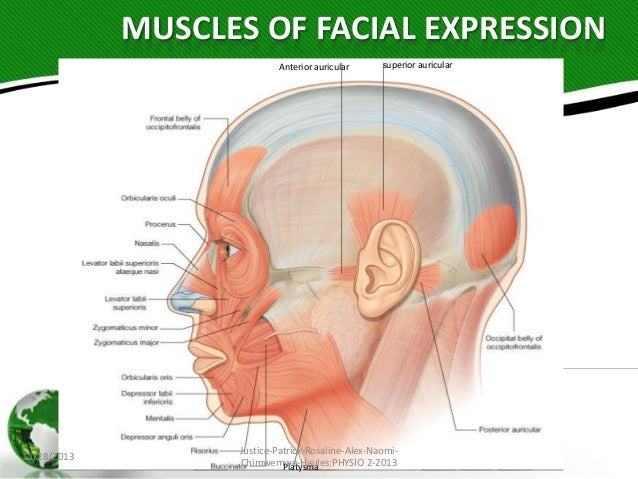
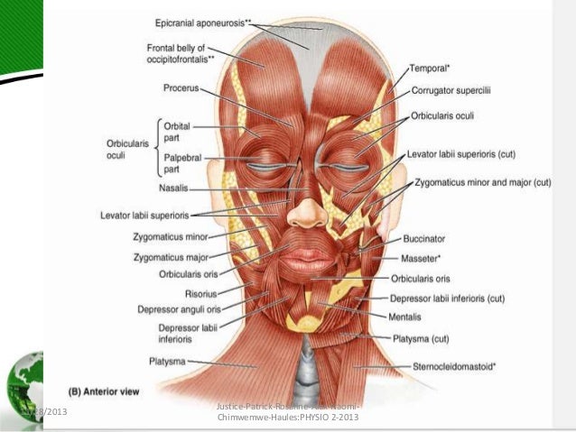
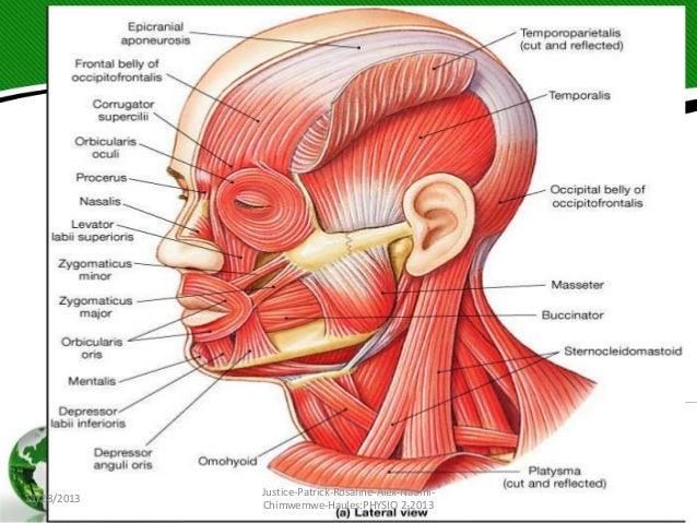
Interactive tutorials on the facial expression muscles, featuring the beautiful diagrams and illustrations of GetBodySmart. Start learning now!
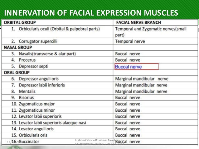
Central facial palsy (colloquially referred to as central seven) is a symptom or finding characterized by paralysis or paresis of the lower half of one side of the face.It usually results from damage to upper motor neurons of the facial nerve.
Chapter 5: Facial sensations & movements. In this chapter, the functions of the trigeminal (CN V) and facial (CN VII) nerves will be discussed.


Facial muscles. The facial muscles are a group of about 20 flat skeletal muscles lying under the facial skin. Most of them originate from the skull or fibrous structures and radiate to the skin through an elastic tendon.

The facial nerve, CN VII, is the seventh paired cranial nerve. In this article, we shall look at the anatomical course of the nerve, and the motor, sensory and parasympathetic functions of its terminal branches.


SYNONYMS: Cranial nerve seven (VII), Nervus facialis COURSE OF FACIAL NERVE Supranuclear pathways 1. Somatomotor cortex: controlling motor component of facial nerve lies in precentral gyrus (Broadmann area 4,6,8) 2.
Pectoralis Major Muscle – Attachment, Action & Innervation. Pectoralis major is a thick, fan-shaped muscle contributing to the thoracobrachial motion.

Each nerve is covered externally by a dense sheath of connective tissue, the epineurium.Underlying this is a layer of flat cells, the perineurium, which forms a complete sleeve around a bundle of axons.

The muscles of facial expression are located in the subcutaneous tissue, originating from bone or fascia, and inserting onto the skin. By contracting, the muscles pull on the skin and exert their effects.

Muscle – Tetrapod musculature: In the living urodeles (newts and salamanders) of the class Amphibia, the axial muscles are most important for propulsion. The limbs of urodeles are quite weak and tend to be carried forward …


